Prompt, Reliable Same-Day Visits Available!

Exotic pets—rabbits, birds, reptiles—are masters at hiding illness. By the time visible symptoms appear, a condition may already be advanced. Ultrasound imaging has transformed how specialists detect and diagnose internal problems in these animals before they become life-threatening.
Unlike dogs and cats, exotic species require a uniquely skilled approach, advanced equipment, and a trained eye that understands their distinct anatomy. This article walks through how board-certified internists use diagnostic ultrasound in exotic pets, what conditions it can uncover, and why finding an experienced exotic pet specialist early can make all the difference for your rabbit, bird, or reptile.
Why Exotic Pets Need Specialized Diagnostic Imaging
Most exotic animals are prey species. Instinct drives them to mask pain and weakness, which means owners rarely notice something is wrong until the disease has progressed significantly. A rabbit may eat normally for weeks while a liver lobe enlarges silently. A bird may appear active and vocal while carrying a reproductive mass. A bearded dragon may bask under its lamp with an intestinal obstruction developing internally.
Standard physical exams—however thorough—have limits. Palpation can detect large abnormalities in some species, but small body cavities, dense musculature, or thick shells in reptiles make internal assessment nearly impossible without imaging.
This is where diagnostic ultrasound becomes essential.
What Is Exotic Pet Ultrasound and How Does It Work?
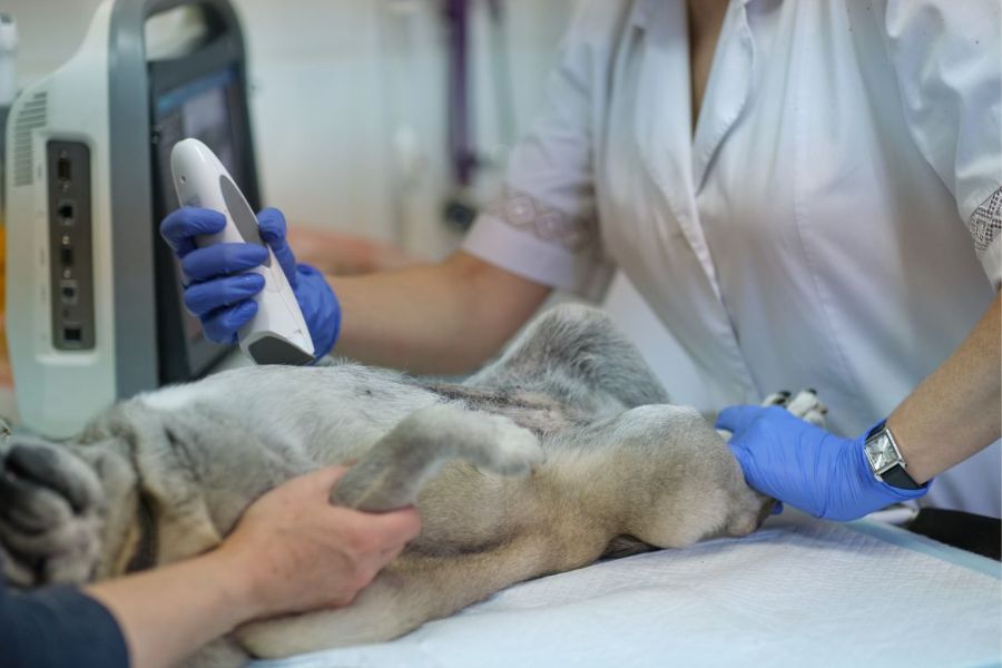
Ultrasound uses high-frequency sound waves to create real-time images of internal organs and tissues. A small probe placed against the skin (or shell, in reptiles) transmits sound pulses that bounce back differently depending on tissue density. The returning echoes are processed into a live image on screen.
Unlike X-rays, ultrasound reveals soft tissue detail with precision—organ texture, fluid accumulation, masses, and even blood flow through vessels (via Doppler mode). For exotic pets, this makes it uniquely suited to evaluating the liver, kidneys, reproductive organs, intestines, bladder, spleen, and heart.
The procedure is typically non-invasive and well-tolerated. Most rabbits and birds require minimal or no sedation. Some reptiles may need light sedation depending on temperament and the area being assessed.
Ultrasound in Rabbits: What Internists Look For
Rabbits are prone to several internal conditions that present with vague outward signs—weight loss, lethargy, reduced appetite, or a slightly rounded abdomen.
Common Findings in Rabbit Ultrasound
- Uterine adenocarcinoma — One of the most common cancers in unspayed female rabbits over age three. Ultrasound detects uterine enlargement, irregular margins, and associated lymph node changes.
- Hepatic coccidiosis or liver lobe enlargement — Infections, abscesses, or cysts in the liver are often asymptomatic until quite advanced.
- Kidney disease — Cysts, mineralization, and size abnormalities in the kidneys can be clearly evaluated.
- GI stasis complications — While stasis is often a clinical diagnosis, ultrasound can rule out obstructive causes and assess gut motility in real time.
- Bladder sludge and uroliths — Calcium deposits and stones appear as hyperechoic (bright) foci with shadowing.
A board-certified internist familiar with rabbit anatomy can differentiate normal cecal shadow from pathological masses—a distinction that requires both experience and species-specific knowledge.
Ultrasound in Birds: A Window Into a Compact Anatomy
Avian anatomy presents unique imaging challenges. The air sac system, keel bone, and small body size demand high-frequency probes and precise technique. Despite this, ultrasound remains one of the most useful tools for avian internal medicine.
Common Findings in Bird Ultrasound
- Reproductive disorders — Egg binding, follicular stasis, and ovarian cysts are among the leading causes of illness in female birds. Ultrasound visualizes follicle size, egg position, and free fluid.
- Hepatomegaly (enlarged liver) — Common in parrots and cockatiels with fatty liver disease, infections, or tumors. Ultrasound assesses liver echogenicity and borders.
- Ascites (fluid accumulation) — Fluid in the coelomic cavity appears clearly on ultrasound and can be sampled under guided visualization.
- Renal masses or kidney enlargement — Avian kidneys are embedded in the synsacrum and difficult to assess otherwise.
- Cardiac evaluation — Echocardiography in birds allows measurement of heart chambers and assessment of function.
If your bird has been showing signs of lethargy, tail bobbing, or abdominal swelling, consulting a veterinarian near you with exotic imaging capability is strongly advised.
Ultrasound in Reptiles: Seeing Through Scales and Shell
Reptile imaging is arguably the most technically demanding. Scutes, scales, and shells alter probe placement. Ectothermic metabolism means organ size and position shift with temperature. Despite this, ultrasound provides critical diagnostic data in chelonians (turtles, tortoises), lizards, and snakes.
Common Findings in Reptile Ultrasound
- Follicular development and reproductive status — Internists use ultrasound to monitor clutch development, detect dystocia, and assess follicular regression.
- Coelomic masses — Abscesses, parasitic granulomas, and neoplasia can develop silently and are clearly identified on imaging.
- Hepatic pathology — Fatty liver disease (hepatic lipidosis) is common in captive reptiles on unbalanced diets. Ultrasound detects abnormal echogenicity and enlargement.
- Urinary and renal disease — Gout and nephrocalcinosis appear as highly echogenic foci in the kidneys.
- Foreign bodies and GI obstruction — Substrate ingestion is common in captive lizards. Ultrasound helps locate obstructions before surgical intervention.
What to Expect During an Exotic Pet Ultrasound Appointment
The appointment typically begins with a full history review and physical exam. Your specialist will discuss findings and recommend imaging based on clinical signs and initial diagnostics.
For the scan itself:
- A small area of skin may be moistened or lightly clipped for better probe contact.
- Your pet is gently positioned, often in dorsal or lateral recumbency.
- The procedure lasts 15–40 minutes, depending on complexity.
- Images are reviewed immediately; a full report follows within 24–48 hours.
In some cases, ultrasound-guided fine needle aspirates or biopsies may be performed during the same appointment, allowing simultaneous tissue sampling.
Why Board Certification Matters
Not all practitioners have the training to interpret exotic pet ultrasound accurately. Board-certified internists complete years of additional residency training in internal medicine, including advanced imaging interpretation. They understand the normal ranges for exotic species, recognize subtle abnormalities, and integrate findings with bloodwork, radiographs, and clinical presentation.
For complex or unclear cases, having a specialist perform and interpret the scan significantly reduces the margin for diagnostic error.
Conclusion: Early Detection Changes Outcomes
For exotic pets, the window between “undetectable” and “advanced disease” can close quickly. Ultrasound imaging, performed by a board-certified internist, gives owners and clinicians a reliable way to find what can’t be seen from the outside. Whether it’s a mass in a rabbit’s uterus, fluid around a parrot’s liver, or follicles in a tortoise, early detection creates more treatment options and better outcomes.
If you’re searching for a veterinarian near you who has experience with exotic species diagnostics, don’t wait for symptoms to worsen. Reach out to a specialist who understands the anatomy, physiology, and needs of your specific pet.
If you’re located in Tranquility, reach out to Tranquility Veterinary Clinic—a practice where compassionate, expert care for every species comes together with a genuine commitment to the well-being of your pet and your peace of mind.
Frequently Asked Questions(FAQs):
1. Is ultrasound safe for rabbits, birds, and reptiles?
A: Ultrasound uses sound waves rather than radiation, making it one of the safest imaging methods available. It is non-invasive, painless, and well-tolerated by most exotic species with minimal or no sedation required.
2. How do I know if my exotic pet needs an ultrasound?
A: Signs such as unexplained weight loss, lethargy, abdominal swelling, changes in droppings, or reproductive symptoms are all reasons to consider imaging. A specialist can determine if an ultrasound is the right diagnostic step based on your pet’s clinical signs.
3. Can a regular vet perform an exotic pet ultrasound?
A: While general practitioners may have ultrasound equipment, exotic species require specialized anatomical knowledge and calibrated technique. A board-certified internist with exotic animal experience provides the most accurate and reliable imaging results.
4. What conditions can ultrasound detect in birds?
A: Ultrasound is particularly useful for diagnosing reproductive disorders (egg binding, follicular stasis), liver disease, coelomic fluid, renal masses, and cardiac abnormalities in pet birds, including parrots, cockatiels, and finches.
5. Does my reptile need sedation for an ultrasound?
A: It depends on the species and temperament. Many lizards and tortoises tolerate ultrasound calmly, while some snakes or stress-prone individuals may benefit from light sedation to ensure accurate imaging and reduce handling stress.
6. How much does an exotic pet ultrasound cost?
A: Costs vary by species, region, and complexity of the scan. A standard abdominal ultrasound typically ranges from $150–$400, with specialist consultations and guided procedures potentially adding to that total. Your clinic can provide an estimate during the consultation.